|

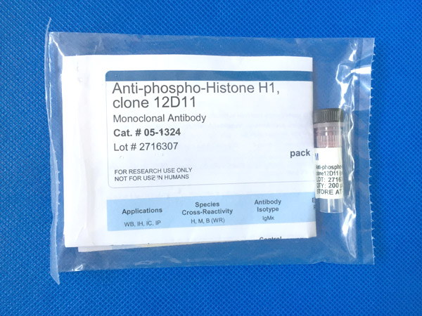
Anti-phospho-Histone H1, clone 12D11
Close
REFERENCES
Growth factors that repress myoblast differentiation sustain phosphorylation of a specific site on histone H1.
Cole, F, et al. (1993) J. Biol. Chem., 268: 1580-5 (1993)
Monoclonal antibody specific for histone H1 phosphorylated by cyclin-dependent kinases: a novel immunohistochemical probe of proliferation and neoplasia
Burstein, D. E., et al (2002) Mod Pathol, 15:705-11 (2002)
Species Reactivity Key Applications Host Format Antibody Type
H, B, M, Vrt IHC, ICC, IP, WB Mouse Culture Supernatant Monoclonal Antibody
Description:
Anti-phospho-Histone H1 Antibody, clone 12D11
Promotional Text:
Special Shipping Offer on Antibodies
100% Performance Guaranteed
Specificity:
Recognizes phosphorylated Histone H1, Mr 32 kDa.
Molecular Weight:
~32 kDa
Immunogen:
The immunogen was KLH-conjugated bovine nuclear extract.
Modifications:
Phosphorylation
Clone:
12D11
Isotype:
IgMκ
Background Information:
Histones are highly conserved proteins that serve as the structural scaffold for the organization of nuclear DNA into chromatin. The four core histones, H2A, H2B, H3, and H4, assemble into an octamer (2 molecules of each). Subsequently, 146 base pairs of DNA are wrapped around the octamer, forming a nucleosome. The linker histone, H1, interacts with linker DNA between nucleosomes and functions in the compaction of chromatin into 30nm chromatin fibers and higher order structures.
View All »
Species Reactivity:
Human
Bovine
Mouse
Vertebrates
Species Reactivity Note:
Human, mouse, bovine. Broad species cross-reactivity is expected, based on sequence homology.
Application Notes:
Immunohistochemistry: This antibody has been reported by an independent laboratory to detect CDK (cyclin dependent kinase) phosphorylated Histone H1 in benign tissue sections.1
Immunocytochemistry:
This antibody has been demonstrated by an outside laboratory to show positive immunostaining for phosphorylated histone H1 in HUSK (human fetal primary myoblasts) cells fixed with 3.7% paraformaldehyde followed by permeabilization with 0.2% Triton X-100.2
Immunoprecipitation:
This antibody has been demonstrated by an outside laboratory to immunoprecipitate phosphorylated histone H1 using solid phase beads bound to anti-mouse IgM.
View All »
Control:
HeLa cell lysate, treated with colcemid.
Quality Assurance:
Western Blot Analysis:
1:1000 dilution of this antibody detected Histone H1 in colcemid-treated HeLa lysate.
Presentation:
Supplied as 200 μL of culture supernatant with 0.05% sodium azide.
Storage Conditions:
Stable for 1 year at -20°C from date of receipt. Upon first thaw, and prior to removing the cap, centrifuge the vial and gently mix the solution. Aliquot into microcentrifuge tubes and store at -20°C. Avoid repeated freeze/thaw cycles, which may damage IgG and affect product performance.
UniProt Number:
P16401
Entrez Gene Number:
NM_005319
Gene Symbol:
H1
H1.5
H1F5
MGC126630
MGC126632
OTTHUMP00000017748
View All »
Alternate Names:
H1 histone family, member 5
Histone H1a
histone 1, H1b
histone cluster 1, H1b
Usage Statement:
Unless otherwise stated in our catalog or other company documentation accompanying the product(s), our products are intended for research use only and are not to be used for any other purpose, which includes but is not limited to, unauthorized commercial uses, |